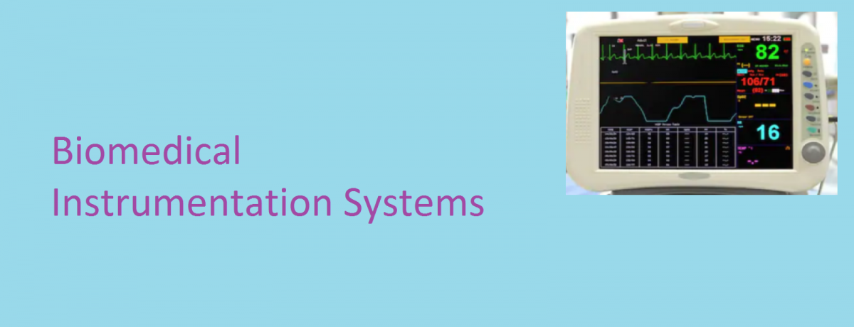
Since the electrical events in the normal heart precede its mechanical function, ECG signals play an important role in diagnosis and monitoring of a wide variety of cardiac abnormalities including myocardial infarction and chamber enlargements. Hence, ECG signal acquisition systems are extensively employed in cardiology, cardiac catheterization laboratories, intensive and cardiac care units, etc.
The electrical activity of the heart can be determined by measuring the potential differences between points on the torso known as surface ECGs. These ECG signals have diagnostically significant frequency content between 0.05 and 150 Hz. To ensure stability of the baseline, a good low frequency response is needed. The instabilities in the baseline recordings originate from changes in the electrode-electrolyte interface at the point of contact of the bioelectrode with the skin. To accurately record fast changes in the ECG signals and distinguish between other interfering signals of biological origin, adequate high frequency response is required. This upper frequency value is a compromise between several factors including limitation of mechanical recording parts of ECG machines using direct writing chart recorders.

To amplify the ECG signals and reject non-biological signals i.e. powerline noise as well as biological interferences e.g. EMG, differential amplifiers with high gains (typically 1000 or 60 dB) with outstanding common mode rejection capabilities must be employed. Typically, common mode rejection ratios (CMMRs) in the range of 80 – 120 dB with 5 kꭥ imbalance between differential amplifier input leads provide a desirable level of environmental and biological noise and artifact rejection in ECG acquisition systems. Additionally, in very noisy environments, it becomes necessary to engage a notch (band-reject) filter centered at 60 Hz or 50 Hz to reduce powerline noise further.
Modern biomedical instruments use instrumentation amplifiers. These amplifiers are advanced versions of differential amplifiers enhanced with many additional desirable characteristics such as very high (input impedance > 100 Mꭥ) to prevent loading the small ECG signals that are to be picked from the skin. The final stages of the ECG amplifier module limit the system’s response (band pass filtering) to the desirable range of frequencies for diagnostic (i.e. 0.05 – 150 Hz) or monitoring (i.e. 0.5 – 40 Hz) purposes. A more limited bandwidth in the monitoring mode provides improved signal to noise ratio and removes ECG baseline drift due to half-cell potentials generated at the electrode/electrolyte interface and motion artifacts. Driven right-leg amplifiers improve the CMRR.
Related: Electrocardiogram
The amplified and sufficiently filtered ECG signals are then applied to display, recording or digitization modules that are computer based.


One thought on “The Instrumentation for Recording ECG Signals”