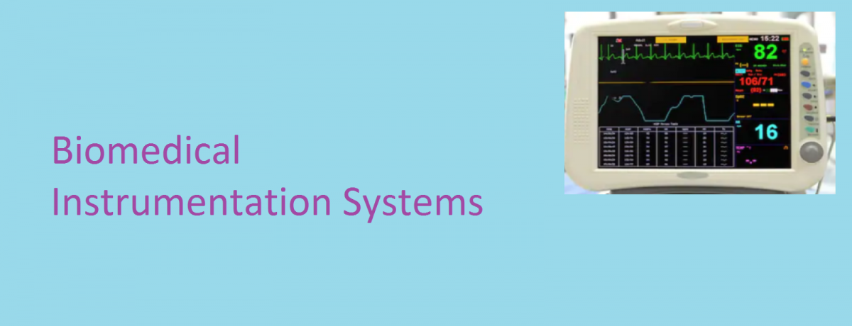
We have different types of biomedical recorders named according to the types of bio signals they capture. Some of these instruments used to record data in biomedical measurements include:
Contents
Electrocardiograph (ECG)
The Electrocardiograph (ECG) is an instrument which records the electrical activity of the heart. Electrical signals from the heart characteristically precede, the normal mechanical function and monitoring of these signals has an important clinical use. ECG instrument provides valuable information about a wide range of cardiac disorders such as the presence of an enlargement (cardiac hypertrophy or presence of an inactive part (infarction). Electrocardiographs are used in coronary care units, catheterization laboratories and for routine diagnostic applications in cardiology.
Vectorcardiograph (VCG)
Vectorcardiograph is the technique of analyzing the electrical activity of the heart by obtaining ECG’s along three axes at right angles to another and displaying any of two of these ECGs as a vector display on an X-Y oscilloscope. The display is known as vectorcardiogram (VCG). The Electrocardiogram displays the electrical potential in any one single axis; the vectorcardiogram displays the same electrical events simultaneously in two perpendicular axes. This gives a vectorial representation of the distribution of electrical potentials generated by the heart, and produces loop type patterns, on the CRT screen.
Phonocardiograph (PCG)
The Phonocardiograph is an instrument used for recording the sounds connected with the pumping action of the heart. These sounds provide an indication of the heart rate and its rhythmicity. They also give useful information regarding effectiveness of blood pumping and valve action.
Diagnostically, heart sounds are very useful. Sounds produced by healthy parts are remarkably identical and abnormal sounds always correlate to specific physical abnormalities. The phonocardiograph provides a recording of the waveforms of the heart sounds. The waveforms are important when it comes to diagnostics and reveal more details than just mere sound itself.
Electroencephalogram (EEG)
This is an instrument for recording the electrical activity of the brain by placing appropriately surface electrodes on the scalp. Monitoring the electroencephalogram has proven to be an effective method of diagnosing many neurological illness and diseases such as epilepsy, tumour, cerebrovascular lesions, ischemia, etc. It is also used in the operating room to facilitate anaesthetics and to establish the integrity of the anesthetized patient’s nervous system.
Electromyography (EMG)
Electromyography is an instrument used for recording the electrical activity of the muscles to determine whether the muscle is contracting or not or for displaying on the CRO & loudspeaker the action potentials spontaneously present in a muscle or those induced by voluntary contractions as a means of detecting the nature and locations of motor unit lesions; or for recording the electrical evoked in a muscle by the stimulation of its nerve. Electromyography is useful for study for several aspects of neuromuscular function, neuromuscular condition, extent of nerve lesion, reflex responses etc.
EMG measurements play a key role in the myoelectric control of prosthetic devices (artificial limbs). Their use here involves picking up EMG signals from the muscles at the terminated nerve endings of the remaining limb and using the signals to activate a mechanical arm. EMG is usually recorded by using surface electrodes or needle electrodes.
You can also read: Cardiac Defibrillators
Electrooculography (EOG)
Electrooculography is the recording of the bio-potentials generated by the eye ball. The EOG potentials are picked by small surface electrodes placed on the skin near the eye. One pair of electrodes is placed above and below the eye to pick up voltages corresponding to vertical movements of the eye ball. Another pair of electrodes is positioned to the left or right of the eye to measure horizontal movement.
Electroretinography (ERG)
It has been established that electrical potentials exists between the cornea and the back of the eye. This potential changes when the eye is illuminated. The process of recording the change in potential when light falls on the eye is called Electroretinography. ERG potentials can be recorded with a pair of electrodes. One of the electrodes is mounted on a contact lens and is direct contact with the cornea. The other electrode is placed on the skin adjacent to the outer cornea of the eye. A reference electrode maybe placed on the forehead.
Learn More On: The Muscle Stimulator
Ballistocardiograph (BCG)
Ballistocardiograph is an instrument that records the movement imparted to the body with each beat of the heart cycle. These movements occur during the ventricular contractions of the heart muscle when the blood is ejected with sufficient force.
Don’t miss out Important Updates, Join Our Newsletter List


2 thoughts on “Biomedical Signals Acquisition Instruments”